Written in a casual, narrative style, this edition has been updated with five new chapters, new case studies, new clinical stories, and discussion questions focusing on ethical, legal, and interperson
This uniquely convenient reference offers important focus on motor dysfunction, hundreds of illustrations and easy-scan charts, patient-teaching points, and expert advice for unusual clinical situatio
The thoroughly revised and updated Second Edition of this text is part of the popular Lippincott Williams & Wilkins MRI Teaching File Series. The book presents 100 actual case studies that cover a
This new 2nd Edition provides revised and updated information on the latest pain procedures being performed in today's practices. The author's present a simple "how-to" approach for the various proce
Physician assistants have speedy access to essential facts in important medical specialty areas. Provides tips for assessing patients from different cultural backgrounds, pediatric and geriatric alert
Dean (neurological surgery, Case Western Reserve U.) and Herbener (radiology, U. Hospitals of Cleveland) correlate color images from the Visible Human Project with radiologic images. Corresponding lin
This chart shows the kidney and surrounding organs, veins and arteries. Illustrations show sectioned left kidney and provide a close-up view of pattern of parenchyma of kidney. The chart also shows ne
As orthopaedic specialty units are being combined with other nursing units or integrated into medical-surgical units, more nurses are frequently unprepared to care for these patients with orthopaedic
This chart shows detailed anatomical view of hair within the skin and of the hair shaft. It illustrates types of scalp hair and hair fiber characteristics. Illustrations show miniaturization of hair f
Shows oral cavity, glands, stomach, liver, pancreas and duodenum. Provides cross sections of wall of the stomach, the jejunum and the colon. Also illustrates arterial supply. Compatibility: BlackBe
Shows cross section of the eye. Also provides lateral and top view of the eye and shows the visual field. Illustrates anterior chamber angle, lens, retina, fundus and the macula lutea. Compatibility:
This chart shows posterior view of the pharynx and shows sagittal section, deep side view, and tonsils. Illustrations provide various views of the larynx: anterior, posterior, side, cut-away side, top
This chart defines temporomandibular joint (TMJ) syndrome. It illustrates the normal jaw closed and open, and the nerves of the temporomandibular region. The chart shows and explains common causes and
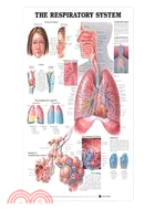
Illustrates the respiratory system from the frontal sinus to the diaphragm. Includes views of the paranasal sinuses, larynx, and bronchopulmonary segments. Also shows the structure of intrapulmonary
Illustrates the respiratory system from the frontal sinus to the diaphragm. Includes views of the paranasal sinuses, larynx, and bronchopulmonary segments. Also shows the structure of intrapulmonary
This chart shows the kidney and surrounding organs, veins and arteries. Illustrations show sectioned left kidney and provide a close-up view of pattern of parenchyma of kidney. The chart also shows n
Classic illustrations by Peter Bachin. Shows anterior, lateral and posterior views of the skeletal system. Also illustrates portion of long bone, auditory ossicles, ligaments of the right hand (dorsa
This chart defines temporomandibular joint (TMJ) syndrome. It illustrates the normal jaw closed and open, and the nerves of the temporomandibular region. The chart shows and explains common causes an